Real Animal Cell Under Microscope - Q Why Do Animal Cells Stay So Small A Gravity The Atlantic / 1 and 100 micrometers and are thus visible only with the aid of a microscope.
Plant and animal cells have their similarities and differences. Cal microscopy is limited in resolution by the wavelength of visible light. In animals and plants each cell is surrounded by a very. The internal structure of cells is studied by transmission electron microscopy, which allows cell organelles to be visualised. What is the method used for preparing an animal cell to look under a.

Plant and animal cells have their similarities and differences.
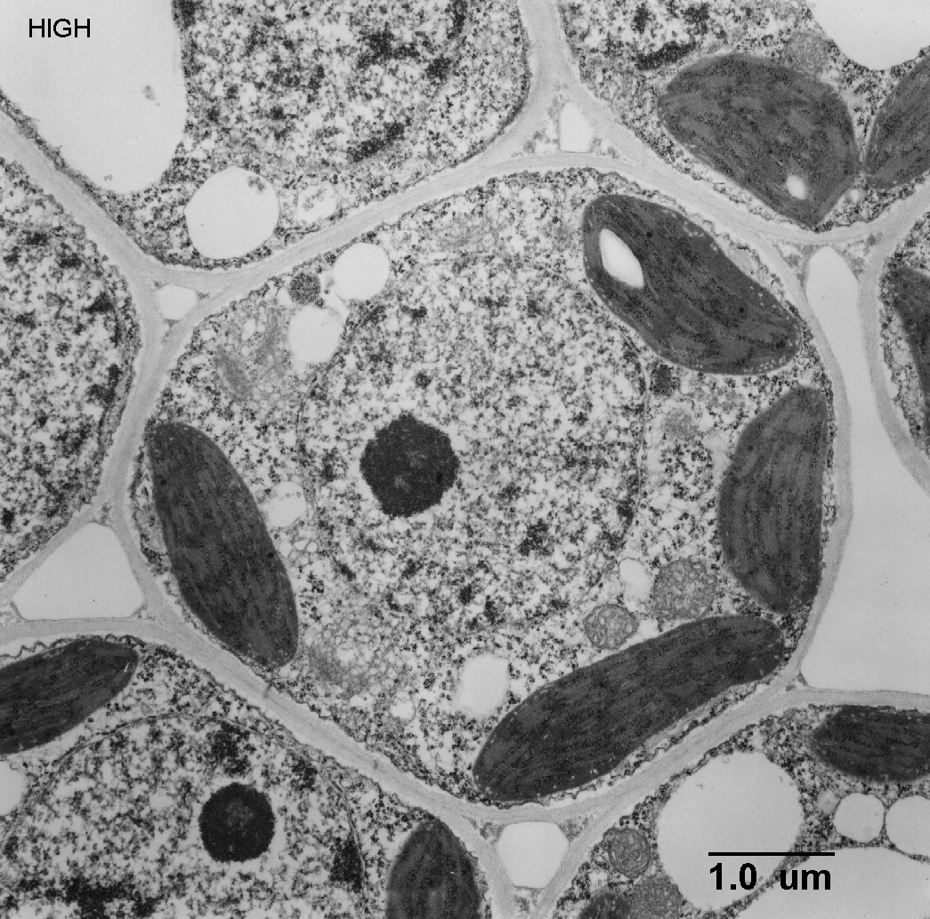
Figure 1.6 shows some actual. Animal cells are typical of the eukaryotic cell type, enclosed by a plasma. Cal microscopy is limited in resolution by the wavelength of visible light. "why do you think the plant cell looks rectangular in shape?" "what . Generalised plant cell as seen with a light microscope. For example, something that you draw as 3cm long, may in fact be 10, 000 times smaller in real life. What are 2 disadvantages of using a light microscope? But, when we view an actual organelle under a microscope, it may look quite different depending on how it was cut into a very thin section to view. Use the equation to work out the real width of the cell in mm: Plant and animal cells have their similarities and differences. In animals and plants each cell is surrounded by a very. Viewing animal cells under a microscope. Figure 1 shows a human cheek cell viewed under a light microscope.
Another kind of electron microscope, a scanning electron microscope. What are 2 disadvantages of using a light microscope? 1 and 100 micrometers and are thus visible only with the aid of a microscope. So it is important to note that what we are drawing is . Animal cells are typical of the eukaryotic cell type, enclosed by a plasma.
Generalised plant cell as seen with a light microscope.
So it is important to note that what we are drawing is . "why do you think the plant cell looks rectangular in shape?" "what . Use the equation to work out the real width of the cell in mm: What is the method used for preparing an animal cell to look under a. For example, something that you draw as 3cm long, may in fact be 10, 000 times smaller in real life. 1 and 100 micrometers and are thus visible only with the aid of a microscope. The internal structure of cells is studied by transmission electron microscopy, which allows cell organelles to be visualised. Another kind of electron microscope, a scanning electron microscope. But, when we view an actual organelle under a microscope, it may look quite different depending on how it was cut into a very thin section to view. Generalised plant cell as seen with a light microscope. In animals and plants each cell is surrounded by a very. Animal cells are typical of the eukaryotic cell type, enclosed by a plasma. Figure 1 shows a human cheek cell viewed under a light microscope.
Figure 1.6 shows some actual. Have students draw a diagram of what they see under the microscope. Viewing animal cells under a microscope. Figure 1 shows a human cheek cell viewed under a light microscope. Generalised plant cell as seen with a light microscope.

Figure 1.6 shows some actual.
Figure 1 shows a human cheek cell viewed under a light microscope. 1 and 100 micrometers and are thus visible only with the aid of a microscope. What are 2 disadvantages of using a light microscope? But, when we view an actual organelle under a microscope, it may look quite different depending on how it was cut into a very thin section to view. Cal microscopy is limited in resolution by the wavelength of visible light. Another kind of electron microscope, a scanning electron microscope. In animals and plants each cell is surrounded by a very. So it is important to note that what we are drawing is . Viewing animal cells under a microscope. Figure 1.6 shows some actual. Use the equation to work out the real width of the cell in mm: The internal structure of cells is studied by transmission electron microscopy, which allows cell organelles to be visualised. Viewing animal cells under a microscope.
Real Animal Cell Under Microscope - Q Why Do Animal Cells Stay So Small A Gravity The Atlantic / 1 and 100 micrometers and are thus visible only with the aid of a microscope.. Use the equation to work out the real width of the cell in mm: Viewing animal cells under a microscope. Figure 1.6 shows some actual. For example, something that you draw as 3cm long, may in fact be 10, 000 times smaller in real life. Viewing animal cells under a microscope.

Post a Comment for "Real Animal Cell Under Microscope - Q Why Do Animal Cells Stay So Small A Gravity The Atlantic / 1 and 100 micrometers and are thus visible only with the aid of a microscope."